×
![Buchcover ISBN 9783967292367]()
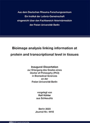
Bioimage analysis linking information at protein and transcriptional level in tissues
von Ralf KöhlerThe optical resolution by human eyes is limited. This is why microscopes were invented, to enable the observation of smaller objects through a specific arrangement of optical components, as well as the investigation of biological processes regarding disease or treatment. Through the microscope’s eye, the camera, images of various tissue stainings can be digitalized and stored on computers. At this time, image interpretation is often performed by the investigator. Manual annotation or counting is a subjective process by the observer, time consuming and therefore perhaps biased. Additional physical limitations increase the chance of misinterpretation regarding aberrations or other distortion artifacts, as well as complexity, which increases by the growing number of available parameters from the same location, notably in spatial gene expression detection. Bioimage analysis, as a newly emerging field in recent years, combines data analysis and imaging methods related to the study of biological processes and has led me to this work, where image data from various microscopy techniques were used to create supervised- and unsupervised bioimage analysis workflows validating biological questions in tissue samples from mouse and human, which were conceptually developed on the MELC system. Thereby, the MELC-system was improved to its overall image quality regarding resolution (laterally 60%, axial 250%), duration of MELCexperiments (reduction of heat and related drying of tissue sample, increase of detectable fluorescence signals) and signal evaluation (artifact removal). The application of system related artifact removal algorithms, such as image registration, illumination correction, image projections or destriping were part of required image pre-processing, which standardized subsequent image segmentation in ilastik and CellProfiler. Classification of found objects or cells benefited from clustering or dimensionality reduction methods simplifying complexity of multi parameter data sets. The application of neighborhood analysis supported the investigation with regard to spatial localizations and interaction. Based on quantitative image analysis workflows developed, including image pre-processing, image segmentation, object identification, classification and representation of the results, a general path from image acquisition to meaningful data evaluation as tool for users could was realized in case of LSM, MELC and LSFM data. Furthermore, the application of MELC and ST linked the information obtained between the protein and transcriptional level. In this way several observations were made, which helped to answer questions in immunology and neuroscience. Segmented LSM images of microglia from aging mice revealed the morphological changes and loss of processes during the course of dementia, using DFT descriptors as a compact way to describe complex shapes. The distribution of the complex compartment of stromal markers in the bone marrow of mice was heterogeneous, which can provide insights into their roles in hematopoiesis and immune cell development. Supervised and unsupervised image analysis workflows identified rare ILC populations in MELC images of mouse tonsils. In addition, their location around vessels and fibronectin fibers was detected by neighborhood analysis. Finally, SARS-CoV-2-induced tissue remodeling was observed in human lung samples from MELC and ST experiments.


